

Brain, nerves, spinal cord, electrical impulses, receptors, effectors, stimuli, response, autonomous, reflex arc.
When you place your hand over a hot flame (say for instance, a candle) and hold it there, what happens? At some point in the near future, you're going to have to take your hand away for it being too hot to handle. This is a motion that requires your central nervous system.
Your brain is connected to almost every part of your body, through a network known as the central nervous system. Within this system, electrical impulses tells parts of your body to move, like the fingers typing up this piece of text. My brain is telling my fingers where to move on a standard keyboard layout, and my fingers are reacting to the nerve impulses to type.
A closer look at this is needed.
Wrinkles in the brain are an evolutionary trait - folds allow more brain matter to fit in the skull without increasing the skull's size. As someone ages, there are more and more folds present.
Much like the other systems in our body, it covers all parts of it. The nerves and endings all react to what the brain tells it to do. This itself is managed by electrical impulses through nerve cells.
The following still is from a video on the reflex arc, with the following details:
Please click on the image to view the video.
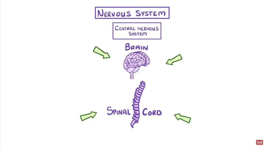
So, back to the candle. Your brain has told your hand to move and place itself just above the candle flame. Inside you, there's an electrical impulse that travels from your brain, down a network of synaptic pathways to the parts of your body that you end up moving (for this instance, your arm and hand).
When it gets too hot, receptorsThe end of a nerve that changes to an electric impulse. in your hand (I'm assuming that's the part you've placed over the flame, and not your arm - hey, don't argue with me!) tell your brain that something is getting hot, and that you need to move your hand out of the way. A signal is sent from the area of your hand to your brain asking for permission to move. The brain accepts defeat and then moves the arm on its behalf, using effectorsThe muscle reacting to the stimuli.. These are the muscle or gland responses that move your arm away from the flame.
The heat from the candle is the stimuli in this instance.
The movement away from the heat is the response your brain will message down to the hand.
This is all done autonomously, and also at lightning speed - I mean, how fast does electricity travel down a wire; ever tried disconnecting your phone from the wire when on charge, turning off the power supply and reconnecting your phone. You get a millisecond of power charge from the wire. The same things happen with your brain and central nervous system.
Often you may find that you lose your balance. This could be because of clumsiness, but could also be an imbalance of the ear. But how does that affect you, and what links it to the central nervous system?
Well, your ear is made up of a series of parts:
This is the visible part of your ear. The ear lobe (the flabby part at the bottom where some of you may have earrings), and the auricle (also known as the pinna). Depending how your genetic makeup is, your ears can have many different shapes and sizes, but they still work the same.
The external auditory canal is the tubular device of the ear that travels inside your head. This is closed off by a tympanic membraneMore commonly known as the eardrum.. The idea of the outer ear is to collect sound waves as they move through the air. They are then guided to the tympanic membrane.
Inside the ear, further than the tympanic membrane, there are a series of three bones - the malleus (hammer), incus (anvil) and stapes (stirrup). Together they form the auditory ossicles.
Inside the inner ear are two functional units - the vestibular apparatus, which contains the sensory organs for your postural equilibrium - this means it helps you stand up straight and maintain your balance. The other part is the snail-shell-like cochlea, which is the sensory organ of hearing. Without this, you wouldn't be able to hear. From there, the ear turns into nerves that link with your brain.
You can have a certain amount of deafness in one or both ears. If you have a hearing deficciency, it can be treated. There are implants you can have put in your ear, and also hearing aids that can help you hear properly.
If you are unable to see the board, please click here.
Brain, forebrain, midbrain, hindbrain, medulla, frontal lobe, parietal lobe, temporal lobe, occipital lobe, neural pathways, cerebellum, cerebral cortex, grey matter, white matter.
The brain is a major part of our body. We still don't know everything about it, but it provides us with life. It helps us move. It controls what we say. It helps us to feel. It interacts with all of our senses, organs and appendages. It's really quite remarkable.
But, how?
The following still is from a video on the brain, with the following details:
Please click on the image to view the video.
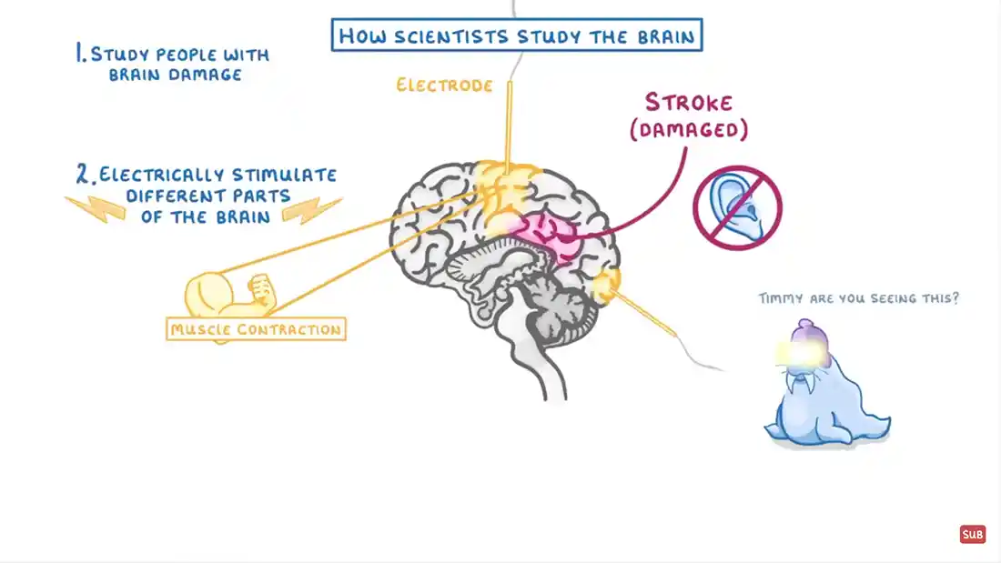
The brain is generally divided into three main sections - the forebrain, the midbrain, and the hindbrain.
The forebrain: this part is at the very front of the brain. It includes the cerebrum, which is made up of both grey matter, and white matter. This grey matter is called the cerebral cortex (cortex is Latin, meaning 'bark'), and the white matter connects two parts of the cortex with a C-shaped structure.
The midbrain: otherwise known as the brainstem, the midbrain comprises a very complex structure of different neuron clusters, neural pathways and other structures. Kind of like a giant mass of motorways joining together, or a collection of rail lines merging. The pons, another part in the midbrain allows us to a range of activities, from tear production, blinking, chewing, focusing vision, balance, hearing and facial expressions.
The medulla is situated at the bottom of the brainstem. This is where the brain meets the spinal cord. It is essential for our survival. The medulla serves to regulate activities such as heart rhythm, breathing, blood flow and oxygen and carbon dioxide levels. It also produces reflexive activities such when we sneeze, swallow a food parcel, or coughing.
The hindbrain: at the back of the brain, the cerebellum is about the size of your fist. It is situated just above the brainstem, and has two sections like the cerebral cortex. The outer part contains neurons, and the inner part interacts with the cerebral cortex. The function of the cerebellum is to coordinate voluntary muscle movements, maintain posture, balance and equilibrium (the state of rest).

The human brain is the fattiest part of our body. Over 60% of it is made of fat out of a total 3kg of weight.
There are four different lobes in the brain. They comprise:
Frontal lobe: by it's name, the frontal lobe is situated at the front of the brain. This part of the brain deals with personality characteristics, decision-making and movement. The frontal lobe also works with our ability to speak, and recognition of smells.
Parietal lobe: in the middle area of the brain is the parietal lobe. This functions to help us identify objects and gives us the ability of hand-to-eye coordination - it lets us be aware of spatial objects around us. The parietal lobe also deals with pain receptors, and the sense of touch in the body. It too helps with the spoken word, this time when we receive spoken word - when someone talks to us, the parietal lobe helps us understand it.
Occipital lobe: the back part of the brain that helps with our vision and the eyes.
Temporal lobe: each side of the brain houses the temporal lobe. This lobe helps us with our short-term memory, speech, muscial rhythm and part of our sense of smell.
There are even more parts of the brain that have not been mentioned, until now. Let's take a look at how the brain is broken down:
The cerebrum is located across the majority of the brain. When you look at a picture of the brain, that shape we all know is what the cerebrum is. But what does it do? Well, it is primarily made up of grey matter around the edges of it, with white matter in the middle. The actual function of the cerebrum allows us to think, learn, keep memory, use our language (or languages if you're lucky enough), deal with our emotions, allow for movement, and perception.
Directly under the cerebrum is the cerebellum. It is about the size of your fist, and it helps to regulate motor behaviours (movement), particularly automatic movements. Not only does it do this, but it allows us to maintain correct posture, balance and also recently has been linked to our understanding and learning.
An interesting note on the cerebellum; although it is only about the size of 10% of the brain, it holds more neurons (nerve cells) than any other part of the brain.
The brainstem is situated at the bottom of the brain, and connects directly to the spinal cord. It acts as a pathway, or relay station between these two areas. It's function is to help us regulate sleep, breathing, body temperature and other functions such as digestion, coughing and sneezing.
The thalamus interacts with relaying sensory information, which contributes to many processes, including perception, attention, timing and movement. The hypothalamus modulates a range of behavioural and physiological functions such as hunger, thirst, body temperature and sexual activity. Through these functions, it integrates information from different parts of the brain, and automatically responds to things like light intensity, smells and also stress.
Part of the brainstem, the pons acts as a bridge. It allows neural pathways to reach their destination. This in turn enables your body to move, and do all the things necessary.
The medulla, also called the medulla oblongata, is a collective name for the medulla, pons and midbrain, and is situated at the lowest point of the brain. It allows the relay of electrical signals to be passed from the spinal cord to the brain and back again.
The spinal cord is explained further down the page.
Nerves, neurons, sensory, relay, motor, brain, movement, senses, spinal cord.
What is a nerve?
Well, it's a strand that allows electronic impulses to be sent through to enable us to move, or touch anything.
There are three main types of nerve cells that are in our body. They include:
While there may be more, they fall under the same category as these.
The following still is from a video on nerves, with the following details:
Please click on the image to view the video.
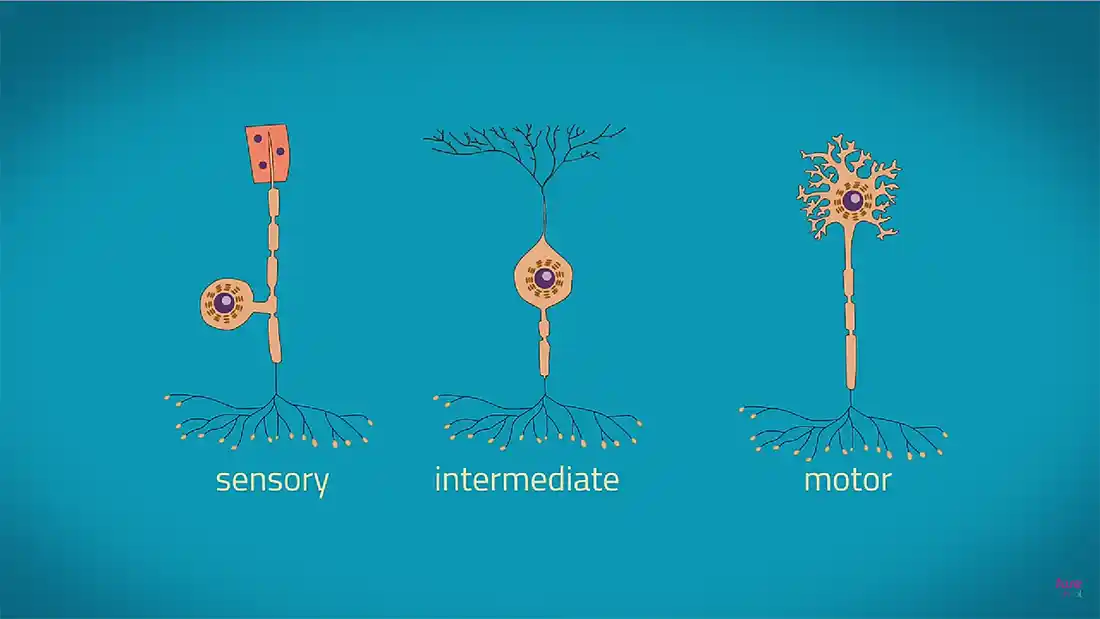
Sensory nerves: they allow us to sense things around us. Like, when we place our hand above a candle, we will feel our hand getting hotter from underneath. They also allow us to react to it (called a reflex action).
Relay nerves: These are between the motor nerve, and the sensory nerves. They are located in the brain and spinal cord, and allow the other nerves to communicate between each other.
Motor nerves: these allow us to move our body. Impulses come from the brain to tell us to move a part of our body - like while I'm typing this, my brain is telling my fingers to touch type the letters on to the document I'm typing it on.

Despite the rest of our body having the capability of being able to regenerate itself to some degree, our nerves/neurons do not have this function, which is often why nerve damage is permanent.
There are several parts of a nerve, including:
The Axon is a strand of the nerve cell that carries information from one end of the nerve cell to the other end of the nerve cell. They are very thin fibres that connect to other neurons (nerves), and also muscle or gland cells.
The myelin sheath is a kind of jacket for the axons in the cell. This keeps it all intact and protects it from any form of intrusion. It's made from fats known as lipids and protein that coat each part of the cell called the axon.
At the end of the cell body are little protrusions, which attach to the other nerve cells or muscles in order to transmit chemical reactions to the next nerve.
Literally just before the information is sent between each nerve cell, it has to travel through the pre-synaptic terminal. These have tiny little gaps that are called synaptic clefts. They allow for transmission of data and information between them to create the chemical reaciton needed.
Usually situated within the middle of the cell, the cell body houses the nucleus of the nerve cell. It works in a similar fashion to a regular cell, like a tissue cell or a muscle cell, and houses other organelles.
A receptor cell is similar to the dendrites in the fact that they connect to other neurons, however, these are closed in, and not free. Being encapsulated means that they allow for more protection.
Rods, cones, accommodation, hyperopia, myopia, eye, sight, looking, iris, pupil, cornea, sclera, macula, optic nerve, retina.
We all need to be able to see, right? So, what do we use? Our eyes. They are the windows to the world for us, and we need to be able to understand them, as well as look after them. So, how do they work?
The eye is a circular (and sometimes oval) ball in our skulls that allow us to see. We base a camera on the same function as the eye. A camera has a moving focal point, which enables the camera to see things up close, as well as far away. Our eyes work in a similar fashion to this.
We can sometimes have conditions or instances where we cannot see properly, and for this, we have treatments in the forms of glasses, contact lenses and corrective laser surgery to fix these issues.

The eye hs 256 unique characteristics. This is better than a fingerprint, which has around 40, and hence why retina scans are more and more popular.
We know these two terms as long sighted and short sighted. But what does that actually mean?
When you're long sighted (hyperopia), you will be able to see things easily that are very far away, but not very well close up.
When you're short sighted (myopia), you will be able to see things easily that are very near, but not very well that are far away.
To correct both of these issues, you can do one of a few things:
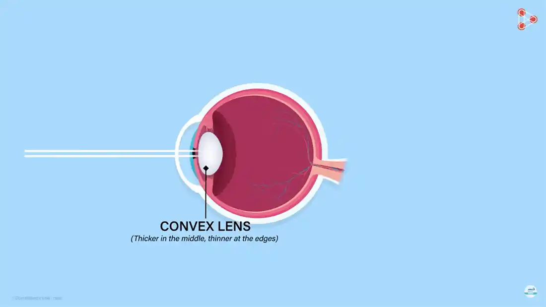
There are several parts to the eye that enable us to see properly. If you look at a diagram of the eye, you will see the following parts:
The very front of the eye that we see, the cornea, is a dome shaped tissue that covers the eye. In theory, you can touch this part, but I wouldn't recommend it. Being at the very front of the eye and being open to the air, it can be irritated by dust or other substances. This is where blinking comes in to remove those substances.
The iris is the part of the eye that changes colour as you grow older. When we are all born, we are born with blue eyes, but depending on the dominant genes in your DNA, this can change. They can be green, brown, grey or a mixture of the three in any form.
The pupil is where the light intensity comes through to the back of the eye. It doesn't have any access to a blood vessel, so it takes its supply of oxygen from the air itself. If the pupil did have a blood vessel go across it, we would see things with a red tint or glow all the time. The pupil changes in size depending on the amount of light in the area, much like the aperture of a camera.
The lens enables us to help focus on an object. More precisely, it allows your eye to focus the amount of light that hits the retina. This then allows the signals to go to the brain with the image we see.
The retina is at the back of the eye. This contains photoreceptors (like the negative of an old camera), which are upside down. That's right, the images we see that are the correct way up, are actually seen upside down at first by the brain. The brain processes them to make them the right way up.
Rods and cones in your eye, more specifically in the retina, are tiny cells that are known as photoreceptors. They allow your brain to see colours, and the amount of light that enters the eye.
Cones are conical in shape, and are made of a protein called photopsin. There are three types of cone, each receptive of a certain colour. Like your television at home, which has a range of colours to be visible using the LED or LCD screen. Your eyes also work in similar fashion.
The cones are separated into the three colours - blue (10%), red (60%) and green (30%). They are responsible for central vision.
Rods are cylindrical in shape, and are also made of a protein, this time called rhodopsin. This allows you to see things in lower light intensity (when it's dusk for example). Although they allow us to see in the darker moments of the day, they do not allow for colour, so often we find it difficult to see in the dark.
Situated at the back of the eye, the optic nerve sends the signal from the eye of the image that we see back to the brain for processing. Once processed, this is then flipped back the right way round, and sent back to the eye to give us our sight and vision. It is lightning quick, as it is pure electrical signalling.
Also situated at the back of the eye, the macula provides us with clear vision. For example, take a projector and a screen. The screen needs to be a certain distance from the projector in order for it to be focal (sharp sighted) and enable us to see clearly. This is what the macula does to enable us to see clearly. It can be given a different focal position (like when we 'stare into space' for example), as your gaze can be changed.
The conjunctiva is located at the front of the eye, above and below the cornea, and is simply a membrane that helps protect the eye. It links to the eyelids so you can close them and open them properly, and is situated on top of the sclera.
The sclera is the part of the eye that is known as the whites. This is the part we see in the mirror everyday when we look at them, and they are usually white in colour. We can get conditions that change their colour, such as conjunctivitis (pink-eye), and can get inflamed.
On the inside, the sclera is not white, it is brown, and has grooves in it, which attaches to the tendons of the eye.
No, it's not funny. Honestly. What it is is a fluid susbtance that fills the eye. It's split into two chambers: one is at the front of the eye, called the anterior chamber. This allows movement of the iris and pupil, and the posterior chamber is at the back in the main 'bulk' of the eye.
This area is also known as the vitreous humour. It helps to maintain the shape of the eye, and gives your vision clarity. It can also absorb a shock to the head, so if for instance, you were to hit a football with your head, it would resonate into the eye and be absorbed. It's like a failsafe system for the head.
The following still is from a video on the eye, with the following details:
Please click on the image to view the video.
Spinal cord, sacral, lumbar, thoracic, cervical, coccygeal, fibrous, vertebra, brainstem, tumours, abcesses, hematoma, fractures.
The spinal cord is a continuation of the brainstem, including a thick bundle of nerves that travel down the inside of your spine. This extends all the way along the spine, and through smaller fibres, is anchored into place at the first Coccygeal vertebra.
The length of the spinal cord in an adult is 40cm, and 2cm wide. Within this cord, it includes:
From this, pairs of the different types of cords emerge from the spine between spaces in it, to go to different parts of the body. This is a vital function of the spinal cord so that it can enable you to do everyday activities you need to perform.
The following still is from a video on the spinal cord, with the following details:
Please click on the image to view the video.
The spinal cord is like the motorway of our central nervous system, with other 'roads' that attach to it to allow impulses to send and receive back to the brain. It is an integral part of the nervous system, and without it we would be paralysed.
Much like the brain, the spinal cord has grey matter, which is situated within the cord flesh. This is a bit like an orange. If you take a look at one, they are encased in the skin (in the spinal cord, this is called the anterior funiculus), and the inside of the orange, which is the part that we eat, is then the grey matter in the spinal cord.
From the spinal cord, four roots appear, which form on either side of the cord. They join together to form the two pairs of cords that emerge and go elsewhere in the body for us to be able to function.
As much as we take our back, our spine and the spinal cord for granted, if we do not look after it, it will deteriorate to a lesser function, and possibly stop altogether. There are a range of disorders that affect the spinal cord, including:
Any of these can be treated, but there are also diseases and conditions that impair the cord further, such as:
While some of the symptoms of these can be reversed, there are no cures for some of them, and they can have permanent damaging effects on your body.
Disclaimer | About Me | Sitemap
Website design by SyntaxHTML.


Blue icons adapted from icons courtesy of Smashicons.com